 |
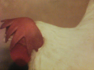
| Figure 1 |
Necrotic
enteris (NE) was first described in chickens in 1961. NE is caused by the
gram-positive bacterium, Clostridium
perfringens. This article discusses a brief review of NE in the literature
and a case report of NE in young commercial pullets in India.
Clostridium perfringens
It is an anaerobic bacterium, i.e. one that grows in the absence of oxygen, and it has the ability to form spores. The spores are small structures highly resistant to environmental stresses. They consist of a tough hard coat that encapsulates the bacterial genetic material and protein necessary for growth. By virtue of forming spores, the bacteria enter a dormant phase when environmental conditions are unfavourable and remain so for as long as necessary. Once present within a chicken house, clostridial spores can remain alive for centuries. Clostridia are capable of producing among the mos potent toxins ever known. It is the toxins that responsible for causing disease. They cause damage to the tissue and destroy red blood cells, thereby reducing the oxygen carrying capacity of the blood. In some cases, they prevent nerves from sending signals to the heart and lungs.
What is necrotic enteritis?
NE is a condition characterised by death or necrosis of the intestinal lining predominantly of the middle and lower small intestine which may be accompanied by necrosis in caeca and liver in some cases. It is transmitted with the ingestion of droppings contaminated soil, water, feed, or litter.
 |
| Figure 2 |
Triggering factors
In healthly chickens, clostridial bacteria normally live harmlessly in the lower gut and are found in the caeca and lower large intestine. The pH and high oxygen content of the healthy small intestine do not support growth of the organisms. For NE to occur, there needs to be a triggering factor that tips the balance in favour of the clostridial bacteria allowing them to proliferate and migrate to the upper small intestine.
Among the known trigger factor are:
- Direct damage to the intestinal lining by coccidial challenge or bacterial overgrowth.
- Feed factors that alter the gut environment like rapeseed, fishmeal, wheat or protein level.
- Immunosuppresion which reduces resistance to gut infections, e.g. CAV, IBD, Marek’s disease, physiological stress.
- Physical factors that damage the gut lining like litter material, a lack of grit or a change in physical feed presentation.
A Case report
In a commercial layer flock of 29 weeks, it was noticed that the peak production standard was no being maintained although morality was within an acceptable range. On investigation, it was foud that the birds were suffering from sub-clinical NE.
Figure 1 shows the changed appearance of the birds comb. Necropsy finding revealed enlarged gas-filled small intestine with the NE observed from the duodenum to the ileum (figure 2). The mucosal surface of the affected area of the intestine was covered with a tan orange pseudo-membrane. This “dirty turkish towel” appeareance is commonly associated with NE. Histopathological examination of the small intestine revealed extensive necrosis of the the villi and infiltration of the lamina proproa with mononuclear cells.
It was also observed that the flock was underweight and each bird was receiving only 207kcal metabolissable energy and 16.8g crude protein daily, compared to the requirements of 290kcal and 18g, respectively. Few birds were showing nasal dishcarge also indicative of mycoplasma infection.
Maize, de-oiled soybean meal, broken rice, sorghum, de-oiled peanut cake, fish and squilla were among the major ingredients of the feed.
The farm records indicated that the farmer had similar problems with previous flocks. For the treatment, it was recommendend that the feed formulation be modified to ensure the birds received adequate levels of energy, protein, lysine, methionine etc. Fishmeal and squilla were removed from the feed, which was supplemented with enzymes and tetramutin (combination of tiamulin and chlortetracycline).
Within 15 days, the flock showed good improvement with regards to the hen-day production, bodyweight etc.
-Dr. Avinash Dhawale,
Ready to cook antibiotics free chicken
ReplyDelete